HEART
Structure
The human heart is a muscular, cone-shaped, hollow organ about the size of a fist (about 12cm in length and 9cm in breadth). The heart is situated behind the sternum, between the lungs in the thoracic cavity. The heart is tilted slightly such that its apex is towards the left side.

The major part of the heart is made up of muscles and is called myocardium. The inner lining of the heart is called the endothelium. The heart is covered by a membrane called pericardium. The pericardium encloses the pericardial cavity that houses the heart.

The human heart is four-chambered. The upper chambers are called the atria or the auricles and the lower two chambers are called the ventricles. The two atria are separated by the interatrial septum. The two ventricles are separated from each other by the interventricular septum. The ventricles have more muscular walls than the auricles.
The right side of the heart is concerned with deoxygenated blood and the left side of the heart with the oxygenated blood. The right auricle opens into the lower right ventricle. This opening is guarded by auriculo-ventricular valve (auriculo ventricular valve). This valve is called the tricuspid valve as it has three flaps. The flaps of the valves are connected to the walls of the ventricle by tendons called the chorda tendinae.

The right ventricle opens to a major artery called the pulmonary artery which takes the deoxygenated blood to the lungs. The junction between the pulmonary artery and the right ventricle is guarded by a semilunar valve. This valve prevents the backflow of blood into the ventricle from the artery.
The left auricle receives oxygenated blood from the left and right pulmonary veins coming from the left and right lung respectively. The left ventricle opens into a major artery called the aorta. The junction between the aorta and the left ventricle is also guarded by semilunar valve that prevents backflow of blood from the aorta into the ventricle.


The function of the heart is to pump blood into the blood vessels to ensure that blood reaches all the parts of the body. This is done by the contraction and relaxation of the chambers of the heart. Contraction is called systole and relaxation is called diastole. The pumping action of heart takes place in a rhythmic pattern.
It consists of three stages:- Auricular systole or ventricular diastole
- Auricular diastole or ventricular systole
- Joint diastole or auricular and ventricular diastole
Auricular Systole or Ventricular Diastole
In the first stage, the auricles or the atria contract with enough pressure to squeeze out all the blood from their cavities into the corresponding ventricles. During this time, the openings from the vena cavae in the right auricle and opening of the pulmonary vein in the left auricles remain closed because of the contraction of the auricles. The semilunar valves guarding the opening of the arteries (aorta and pulmonary artery) are also closed. The auriculo ventricular valves remain open letting blood flow under pressure from the auricles to the ventricles. During this stage, auricles contract and ventricles remain relaxed. This stage is therefore called auricular systole or ventricular diastole.
Auricular Diastole or Ventricular Systole
In the second stage, the auricles relax (diastole). Just as they relax, the flaps of the auriculo ventricular valves snap close. This makes the sound heard as the first heart beat. Once the auriculo ventricular valve is closed, the ventricle contracts (systole). The semilunar valves open as the blood rushes into the arteries from the ventricles.
Joint Diastole or Auricular and Ventricular Diastole
In the last stage, the ventricles start to relax (diastole). This gradually brings down the pressure and at the same time the semilunar valves snap shut to prevent backflow of blood into the heart as the pressure has come down. This produces the second heart beat sound. With the closure of the semilunar valves the ventricles relax further and the auriculo ventricular valves open. Thus, at this stage, both auricles and ventricles are in diastole and hence, the stage is called joint diastole.
The occurrence of the periodic series of events during one heartbeat is called a cardiac cycle. During one heartbeat, there are two heart sounds - 'lub' and 'dub'. 'lub' is the first sound and 'dub' is the second sound.If the heart is removed from a living mammal and placed in a well oxygenated salt solution maintained at 37oC, the heart continues to function though at a slower rate.

CONTROL OF HEART FUNCTION
Position of the Sino-atrial and Atrio-ventricular Nodes
There are certain points in the walls of the heart called the nodes. A node is a collection of muscles. There are two major nodes called the sinoatrial node (Sino-atrial ventricular node) and atrioventricular node (atrio ventricular node). Sino-atrial ventricular node is present at the junction of the vena cavae and the right auricle. It is the initiator of the contraction of heart by making the auricles contract. This is the auricular systole. This impulse is generated and spread by the presence of electrochemical gradient. The impulse is carried to atrio ventricular node that is situated almost at the junction of the auricles and the ventricles. It then transmits it to the other nerves of the heart. The atrio ventricular node is connected to a set of modified cardiac fibres called the 'bundle of His'. From the 'bundle of His' arise various other fine fibres that form a network of fibres running all over the ventricles. These fine fibres are called the Purkinje fibres. Thus, the Sino-atrial ventricular node is called the pacemaker.
In case of failure of the pacemaker, it can be replaced by an artificial battery operated pacemaker.THE HEART BEAT
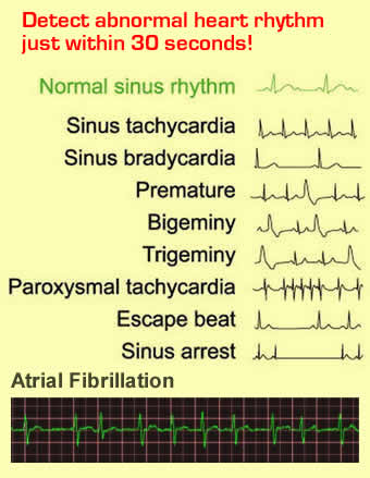
The heart beat results in electropotential differences spreading over the heart muscles. These follow a specific pattern. Any change in the pattern indicates an abnormality. The pattern can be recorded using an electrocardiograph. The recorded pattern is called the electrocardiogram (ECG). This is an important diagnostic tool in detecting any defect in the structure and functioning of the heart.

The heart rate fast or slow, is controlled by the brain and is therefore called the nervous control The brain has two sites, cardiac accelerator site and the cardiac inhibitory site. The nerves run from these sites in the brain to the different nodes of the heart.
Heart Rate and Pulse Rate
The number of times the heart beats per minute is called heart rate. For a normal adult human it is about 72 times per minute. For infants, it is a higher figure. It also goes up during physical exercise.The contractions and relaxations that the heart chambers undergo are felt all along the arteries. If a finger is kept at a spot where an artery runs close to the surface, the rhythmic movement can be felt. This is called the pulse. It is found to be much the same as the heart rate. The number of pulses per minute is called the pulse rate.
There is also hormonal control over the heart rate. The common hormones known to increase the heart rate are the adrenaline and thyroxine. Adrenalin is secreted by the adrenal glands and thyroxine by the thyroid gland.DISEASES AND DISORDERS OF THE CIRCULATORY SYSTEM
Anaemia
The condition in which a number of RBCs per cubic mm being less than normal is called anaemia. It may be due to poor diet or due to excessive bleeding.Sickle-celled Anaemia
It is a congenital disorder where the red blood cells are sicke-celled. This reduces their oxygen-carrying capacity. It is fatal disorder.Arterio-sclerosis
It is the hardening of arteries. High levels of blood cholesterol leads to deposition of cholesterol on the walls of the blood vessels. This makes the arterial walls lose their elasticity and result in their hardening.Hypertension
The arterial blood pressure becomes very high because the stiff walls cannot regulate the pressure changes. This is called hypertension or high blood pressure.Hypotension or Low Blood Pressure
It is the low arterial blood pressure. It may be due to anaemia or malfunctioning of the heart pump.Thrombosis
It is the clot formation in the blood vessels which block the heart. Deposition of cholesterol also leads to thrombosis. The narrowing of blood vessels disrupts the secretion of prostacyclin. Prostacyclin prevents the sticking together of platelets. In the absence of this, the coagulated platelets get deposited in the walls and form the clot or the thrombus. This blocks the blood vessel.P/S: hari ni belajar pasal ni semua.. rasa best sangat2 and dah berinpian nk jadi DOKTOR... teruskan usaha kepada semua..


No comments:
Post a Comment